Coronal CT scan of right eye showing the fish hook.


Right eye CT scan image showing periorbital soft tissue swelling mainly.
Ocular Motility
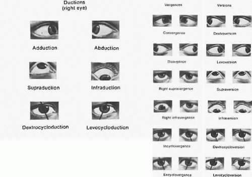
Ocular Motility
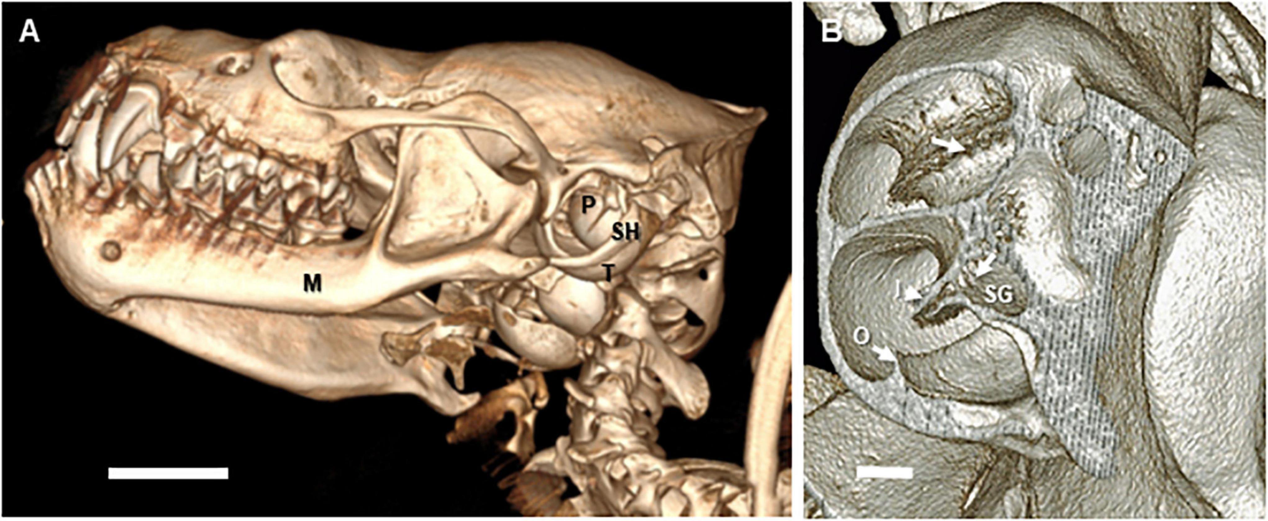
Frontiers Functional Analyses of Peripheral Auditory System Adaptations for Echolocation in Air vs. Water

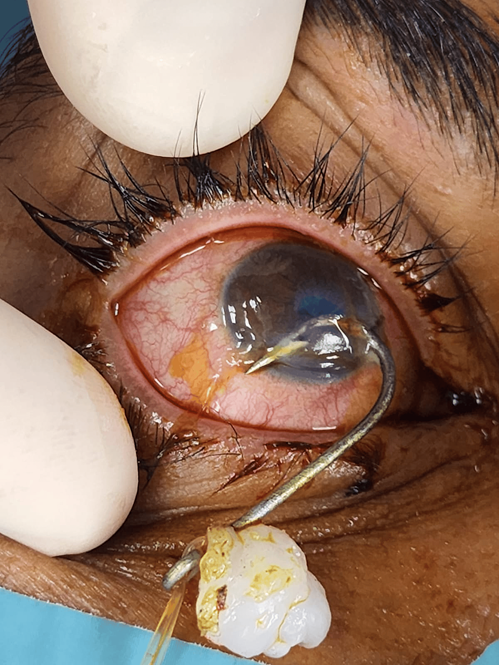
Photograph showing the barbed fish hook penetrating the limbus at 9
Coronal CT scan of right eye showing the fish hook.

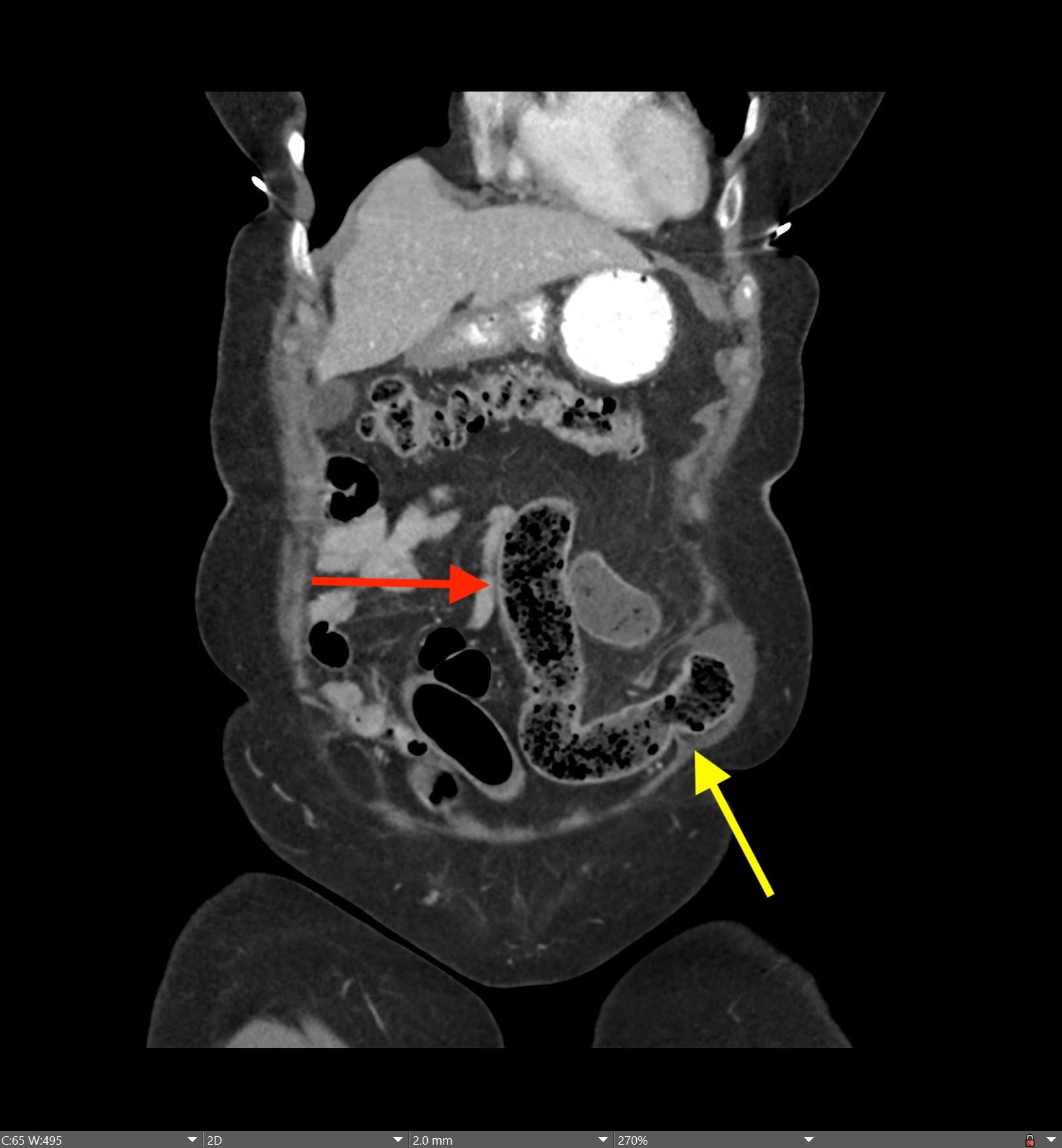
Axial (a) and coronal (b) views of a post eye sparing surgery CT scan

Fish hook penetrating right eye near limbus.(Reproduced from, Sure

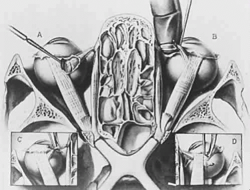
Neurovascular Organization and Assembly of the Face

Handbook Of Ocular Trauma by oculartraumahandbook - Issuu

Swapan SAMANTA, Professor, MBBS (Hons), MS (Ophthalmic Surgery), CCEH(ICEH, London)
Swapan SAMANTA, Professor, MBBS (Hons), MS (Ophthalmic Surgery), CCEH(ICEH, London)
Case of the Month: Department of Radiology: Feinberg School of Medicine
Cureus, Penetrating Ocular Fish-Hook Injury

View Image